Lars Bannow and Benedikt P. Klein awarded the dissertation prize of Philipps-Universität Marburg
Congratulation to Dr. Lars Bannow and Dr. Benedikt P. Klein, PhD-students of the GRK 1782 (SW Koch) and the SFB project A4 (Gottfried), respectively, for being awarded a prize by Philipps-Universität Marburg for their excellent dissertations presented in 2019.
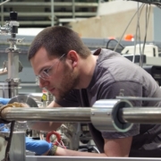
Lars Bannow, GRK 1782 (SW Koch) Foto: Paul Ndimande
In the thesis of Dr. Bannow with the title “Optical and Electronic Properties of Semiconductor Materials”, he investigates optical and electronic properties of novel semiconductor materials such as Ga(AsBi), In(AsBi) and the methylammonium (MA) perovskite MAPbI3. Using a combination of density functional theory calculation and semiconductor Bloch equations, a precise prediction of opto-electronic properties was possible at a minimum of experimental data. The examined materials are promising options for the fabrication of more efficient laser diodes and more economic solar cells.
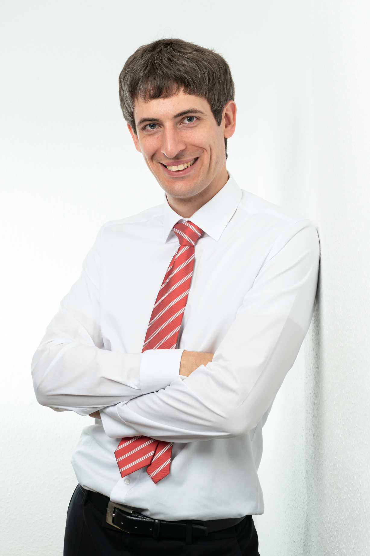
Benedikt Klein, A4 (Gottfried)
In his thesis “The Surface Chemical Bond of Non-alternant Aromatic Molecules on Metal Surfaces”, Dr. Klein explores interfaces between model organic semiconductors and metals. He compares π-electron systems with alternant and non-alternant topologies and finds that the non-alternant topology leads to much stronger interfacial interactions. These studies pave the way to novel organic semiconductors with tailored properties and provide important insight into the bonding of non-alternant defect structures in graphene with metals.
See the news release of the Philipps-Universität Marburg for details of the event.